
Facilities
The Maryland Neuroimaging Center (MNC) is the home for neuroimaging research at the University of Maryland, adjacent to the main College Park campus. MNC is housed in a spacious facility occupying approximately 8000 square feet in the Gudelsky Building, including main reception area, office suites, workspace, conference room, MEG facility, and MRI facilities. It also includes a behavioral testing suite, mock scanner room, and training and classroom space, providing sufficient work areas for multiple labs to streamline the research activities inside the building. The MRI reception area includes the space for the dressing/changing room, interview/testing room, mock scanner room, waiting room, and storage closet. This setup ensures that subject confidentiality is preserved and provides a comfortable waiting area for subjects and their family members.
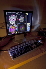
The environment-controlled equipment room and scanner room are used to host major equipment of the MRI system, whereas the fence-protected chiller system for the super conductive magnet is located outside the building. The scanner room is equipped with air-pressure door, RF-shielded window, and LED-powered optical lighting system, whereas the control room provides seating for up to 4 people in addition to the scanning operator. Besides the MR scanner console and its satellite image station, the control room hosts most of the fMRI peripheral equipment, such as fMRI console, stimulation computers, biological recording system, head motion tracking system, eye tracking computer, auditory delivery system, and the equipment for simultaneous EEG and fMRI, etc. First-aids kit, prescription lens kit, and MR-safety screening devices, such as metal wand detector, are also located in the control room. In case of emergencies, an MR-compatible gurney is always standing by in the scanner room, and an automatic external defibrillator (AED) package is available outside the control room door in the hallway.
- MRI
The centerpiece of the facility is a state-of-the-art Siemens 3 Tesla Magnetom PRISMA Fit MRI scanner, equipped with 32 channel head coil and a 64 channel head/neck coil for patient comfort and outstanding signal-to-noise and acquisition speed. - Mock Scanner
Our MRI simulator looks, feels, and sounds like the real thing. It is especially useful for preparing special populations, such as pediatric participants, for their experience in the real scanner, making data acquisition more successful. - MEG
The Yokogawa/KIT/Ricoh Company Magnetoencephalography (MEG) device allows fully non-invasive measurements of neuronal activity in the brain, by recording magnetic fields at 160 different sites around the head. MEG provides exquisite timing resolution and allows for relatively precise localization of many evoked brain responses.